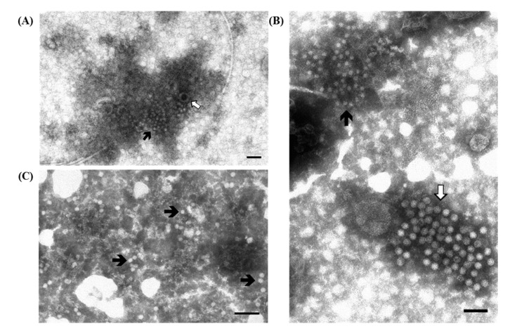
Figure 2.
Electron micrographs of ultracentrifuged fecal contents from pigs with post-weaning enteritis. Small 18–20 nm virions resembling circovirus ( ) are grouped together with (A) a few partially destroyed and empty rotavirus particles (⇧) and (B) a large group of enterovirus-like particles (⇧). Immuno-aggregation electron microscopy preparation with convalescent sera diluted 1:40; (C) IAEM negative control. Several circovirus particles (
 ) are visible, singularly distributed or in small groups. Negative staining (2% sodium phosphotungstate). TEM Philips CM10, 80 kV. Bar = 100 nm.

