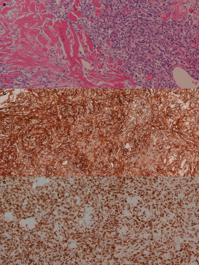
Fig. 4.

Microscopically, the omental tumor was characterized by a cellular proliferation of spindle-shaped cells with no remarkable atypia in a marked collagenous matrix (a, H–E stain ×100). Immunohistochemical test showed the tumor was positive for CD34 (b, ×100) and STAT6 (c, ×100).
