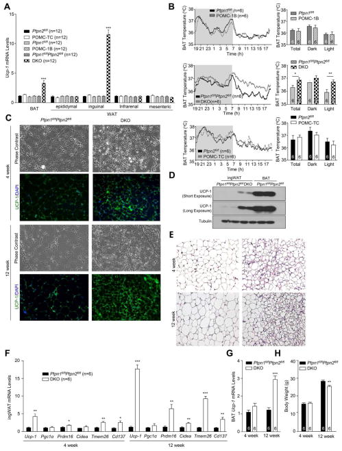
Figure 4. Increased browning in DKO mice.
A) Ucp-1 gene expression in BAT and WAT depots extracted from 12 week-old POMC-1B, POMC-TC, DKO and floxed control mice. b) Interscapular BAT temperatures. c–e) Histology (H&E), immunoblotting and UCP-1 immunohistochemistry in inguinal WAT (IngWAT). f–g) Browning gene expression in ingWAT or BAT and h) body weights. Data are means ± SEM and are representative of 3 independent experiments; significance determined using a) one-way, or, f–g) two-way ANOVA.

