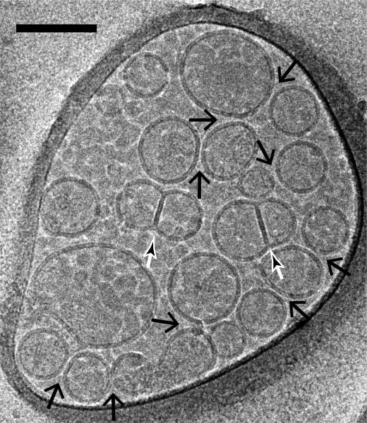
Fig. 4.
Imaging of vesicle-vesicle morphologies by cryo-electron microscopy. Shown are mixtures of v- (with reconstituted synaptobrevin-2) and t-vesicles (with reconstituted syntaxin-1A and SNAP-25A), which were imaged in the holes of the substrate carbon film, visible as the darker areas in the image, in conditions that clearly show the lipid bilayers. Point contacts, (i.e. appositions with small (1-5 nm) separation between vesicles but without merger or deformation of membranes) between docked vesicles were observed (large black arrows) along with hemifused diaphragms (small white/black arrows). The scale bar is 100 nm.

