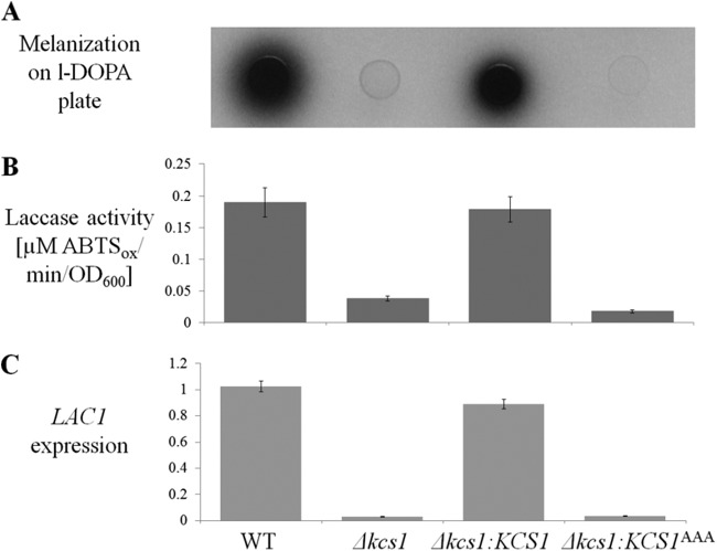
FIG 3 .
Melanization, extracellular laccase activity, and LAC1 gene expression are reduced in the Δkcs1 and kinase-dead Δkcs1::KCS1AAA strains. (A) Cumulative melanization in the WT, Δkcs1, Δkcs1::KCS1AAA, and Δkcs1::KCS1 strains was compared by spotting similar numbers of cells (5 µl; OD600 = 3) onto MM plates lacking glucose (laccase inducing) and containing the laccase substrate l-DOPA, and incubating them for 3 days at 30°C. (B) Extracellular laccase activity was quantified spectrophotometrically by measuring the oxidation of the laccase substrate ABTS (ABTSox) by fungal cells following 4 h of growth in MM broth without glucose at 30°C. (C) LAC1 gene expression was quantified in cultures grown for 5 h in MM broth without glucose by real-time PCR. ACT1 was used for normalization of expression. The differences between different strains in panels B and C are statistically significant (P < 0.05), as determined by Student’s t test.

