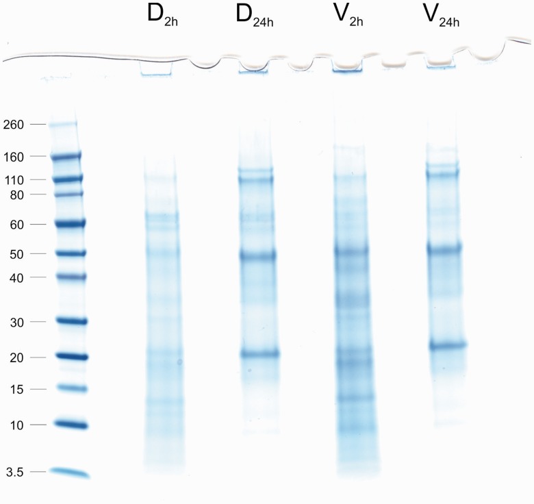
Fig. 2.—
PAGE analysis of M. venosa shell matrices. Lanes D2h (dorsal, 2 h of sodium hypochlorite treatment) and V2h (ventral, 2 h of sodium hypochlorite treatment) show matrix extracted after a 2-h treatment of entire shells with sodium hypochlorite. Lanes D24h and V24h show matrix extracted after an additional treatment of powdered shells for 24 h. The molecular weight of marker proteins is shown in kDa on the left.

