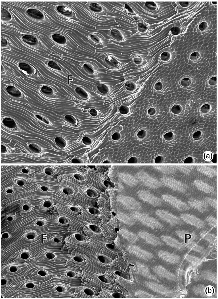
Fig. 6.—
SEM images of punctae in the shell of M. venosa. (a) Punctae in a broken valve of M. venosa (view from inside of shell) with fibrous structure (F) of secondary shell layer clearly visible. (b) Punctae in a broken shell of M. venosa (view from outside of shell) at the interface of the primary (P, right side of image) and fibrous secondary layers (F, left side of image).

