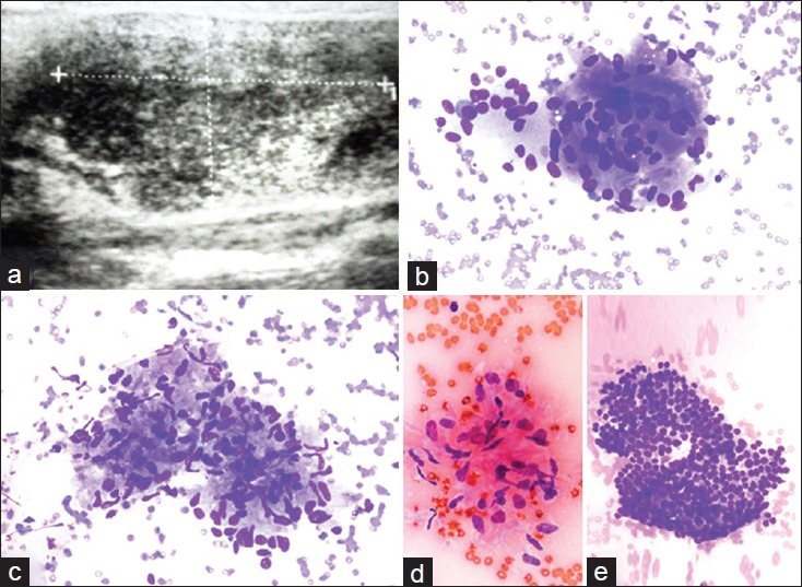
Figure 2.

A subacute granulomatous thyroiditis case with a sonographic image of a poorly defined hypoechoic area. (a) Ultrasound appearance with a discrete area (case no. 19). The cytologic features of the case included the following: (b) Multinucleated giant cell (Diff Quik®, ×400), (c) an aggregate of epitheliod histiocytes consistent with granuloma (Diff Quik®, ×400), (d) a cluster of epithelioid histiocytes (Papanicolau stain × 400) and (e) a group of proliferated follicular cells (Diff Quik®, ×400)
