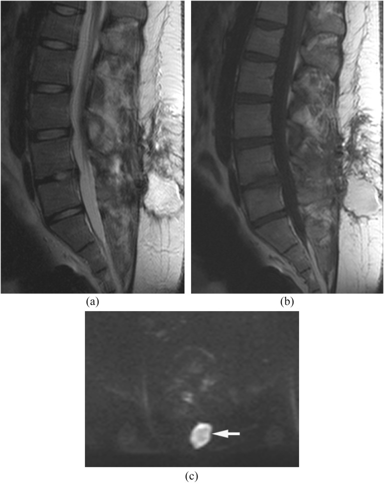
Figure 12.
Post-operative haematoma. (a, b) Sagittal T2 (a) and T1 weighted images (b) show a hyperintense round lesion at the post-operative site. (c) Diffusion-weighted imaging shows hyperintense signal in the lesion (arrow) with decreased apparent diffusion coefficient (not shown) similar to an abscess. A few white blood cells with blood products were proven by drainage.

