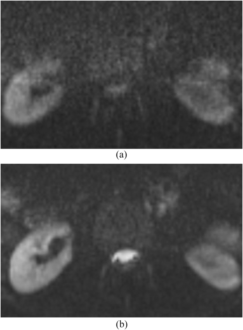
Figure 13.
Diffusion-weighted imaging (DWI) with b-values of 500 vs 1000 s mm−2. (a) We routinely use DWI in spine protocols with a b-value of 1000 s mm−2 for the evaluation of abscess and pus collection. (b) DWI with a b-value of 500 s mm−2 shows partially hyperintense normal cerebrospinal fluid in the spinal canal, which is difficult to differentiate from pus collection. DWI use with low or intermediate b-value is not adequate for this purpose since the signal is more influenced by T2 effect and intravoxel incoherent motion.

