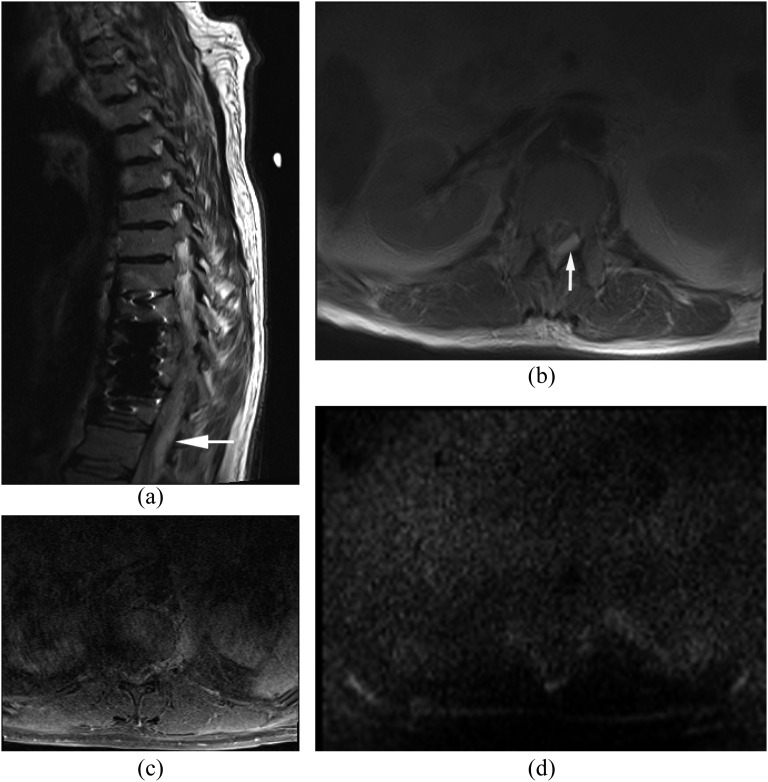
Figure 14.
Susceptibility artefacts. (a, b) Sagittal (a) and axial (b) T2 weighted images show a hyperintense lesion in the epidural space posterolaterally (arrows) with relatively mild susceptibility artefacts from a fusion device. (c) Axial post-contrast T1 weighted image with fat saturation is difficult to detect the epidural abscess owing to motion artefacts. (d) Diffusion-weighted imaging shows no apparent hyperintensity suggestive of the epidural abscess owing to susceptibility artefacts from the fusion device.

