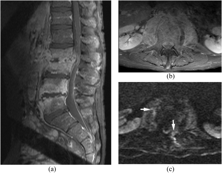
Figure 7.
Candidiasis. Epidural and psoas infections with spondylodiscitis. Candida was proven by biopsy. (a, b) Sagittal (a) and axial (b) post-contrast T1 weighted image with fat saturation show spondylodiscitis at L2/3 and L4/5 with enhancing psoas and epidural lesions. (c) Diffusion-weighted imaging demonstrates hyperintensity in the epidural and psoas lesions suggestive of small pus collections (arrows).

