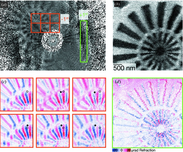
Figure 5.
Holographic STXM. (a) A single holographic image of the Siemens star, illuminated by the −1st-order focus. (b) The horizontal centre-of-mass STXM image if the full detector is used. The images in (c) correspond to the different orange ROIs, while (d) corresponds to the green ROI. The blue-to-red colour map quantifies the refraction angle proportional to differential phase contrast. For a discussion of the circles in (c), see the text. The STXM scan consists of  images, each illuminated for 10 ms, and a field of view of
  µm.
µm.

