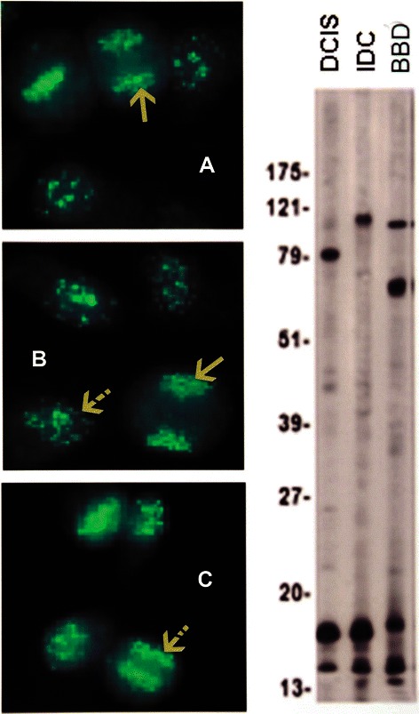
Figure 4.

Anti-centromere antibodies are shown in A, DCIS, B, IDC, and C, BBD. The solid arrows in A and B indicate the fluorescent chromosomes aligned in telophase; the striped arrow in B points to a resting interphase cell, while the arrow in C shows the fluorescent centromeres aligned in metaphase. The immunoblots show the presence of an 80 kDa IgG band corresponding to CENP-B in a DCIS serum.
