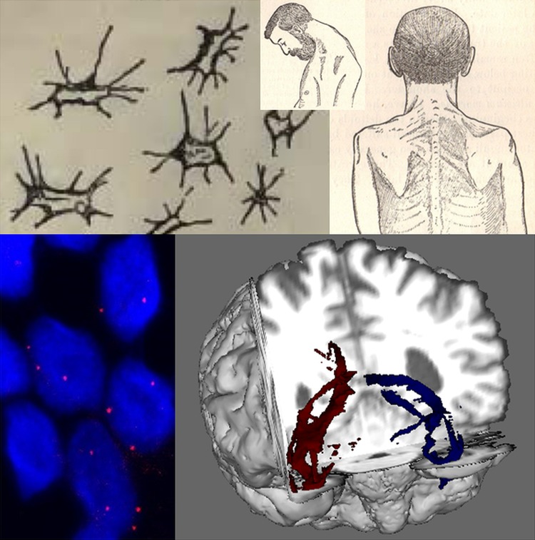
Figure 1.
Developments in cellular and clinical probes for amyotrophic lateral sclerosis (ALS) over 130 years. Lockhart Clarke's hand-drawn atrophied anterior horn cells (top left) are contrasted with RNA foci (below, red dots) visualised within cortical neurons (nuclei, ∼5–10 µ diameter, stained blue with DAPI) differentiated from induced pluripotent stem cells derived from patient with ALS fibroblasts. Gowers’ textbook contained detailed illustrations of a classical ‘flail arm’ variant of ALS with a head drop (top middle and right), contrasted with the white matter tractography of diffusion tensor MRI (bottom right, temporal lobe projection tracts shown in a cutaway coronal plane from the front). Cortical neuron image provided courtesy of Professor Kevin Talbot, University of Oxford.

