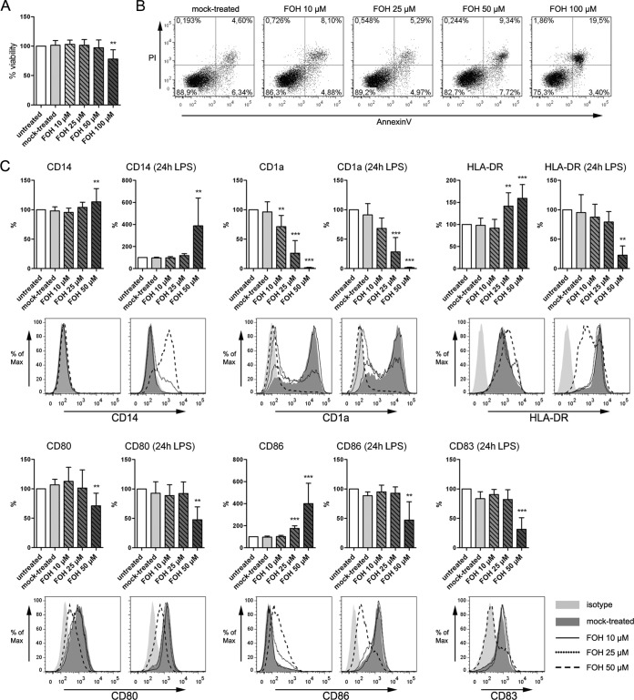
FIG 2 .
Impact of FOH on the differentiation process from monocytes to iDC. iDC generated in the presence of FOH at concentrations ranging from 10 µM to 50 µM showed no impairment of viability but alteration in surface marker expression important for maturation (CD83) and antigen presentation (CD1d, CD80, CD86, and HLA-DR). (A) Quantitative analysis of viability of monocyte-derived dendritic cells (iDC) after differentiation in the presence of FOH. (B) Representative scatter plots showing the distribution of annexin V and PI staining for iDC generated in the presence of FOH. (C) The immunophenotype of iDC generated in the presence of FOH was evaluated. For each surface marker (CD14, CD1a, HLA-DR, CD80, CD83, and CD86), quantitative analysis and a representative histogram profile after 6 days of differentiation (left) and additional 24-h stimulation with LPS (right) are shown. Quantitative data are means and SD from at least 4 independent experiments and normalized to a basal level of untreated DC (white bar) (*, P < 0.05; **, P < 0.01; ***, P < 0.001).

