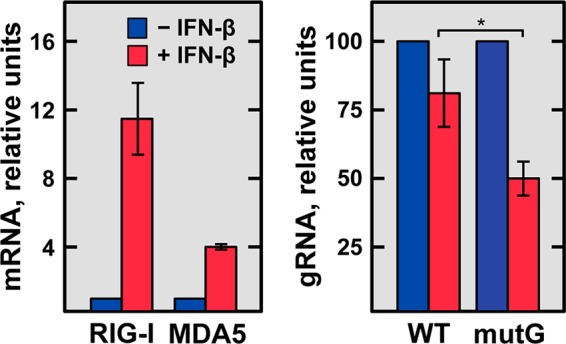
FIG 11 .

IFN-β sensitivity of rTGEV-WT and rTGEV-mutG viruses. Confluent ST cells were untreated (blue) or treated (red) with 4 ng/106 cells of porcine IFN-β (Kingfisher Biotech) for 16 h and then noninfected or infected with either rTGEV-WT (WT) or rTGEV-mutG (mutG) at an MOI of 3. At 8 hpi, total RNA was extracted, and the levels of viral genomic RNA (gRNA) were quantified by RT-qPCR and expressed relative to those in IFN-β-untreated and infected cells (right). As a control of the IFN-β treatment, the MDA5 and RIG-I mRNA levels were quantified by RT-qPCR in noninfected cells, untreated or treated with IFN-β (left). GUSB mRNA levels were used as an endogenous control in all cases. Error bars indicate the standard deviations from three independent experiments. *, P < 0.05.
