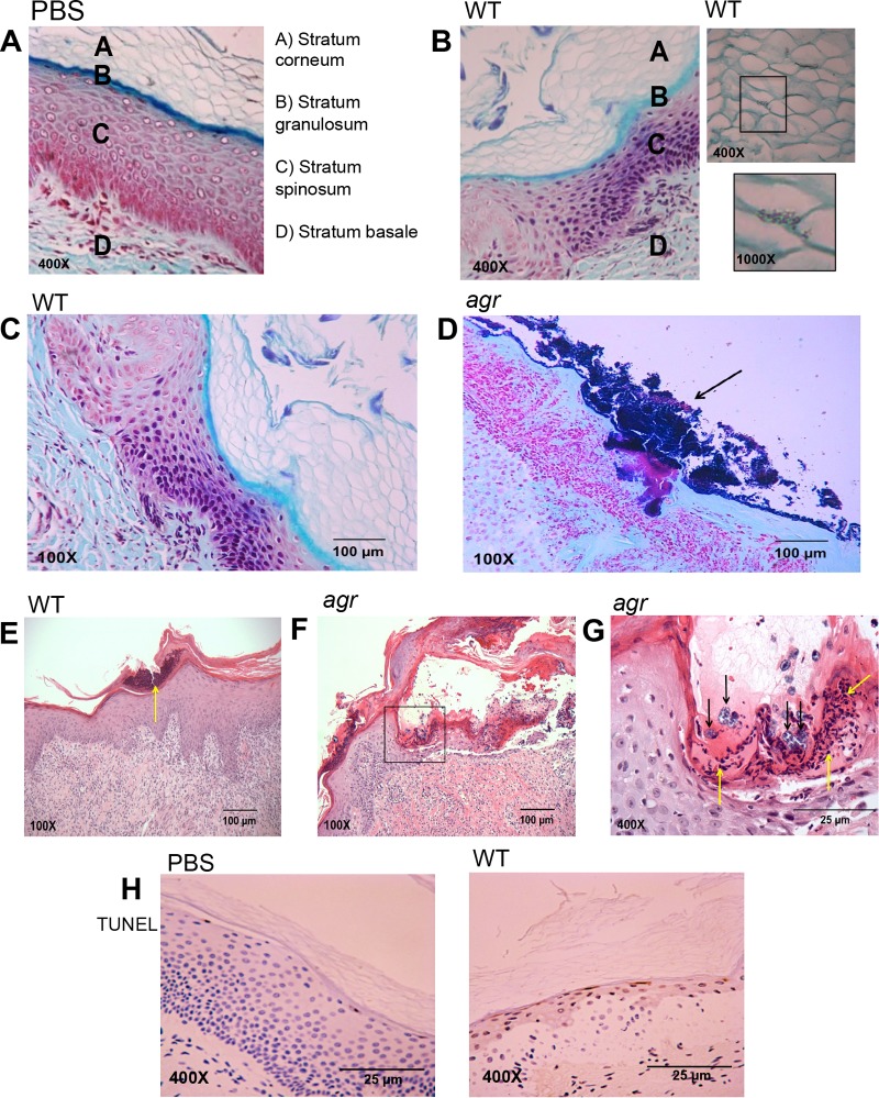
FIG 4 .
Histology of WT S. aureus USA300 and agr mutant infection (108 CFU applied in 10 μl of PBS) of human skin grafts maintained on SCID mice at 72 h postinfection. (A to D) Gram staining with trichrome counterstaining of PBS control (A) or S. aureus infection (B to D). (B) WT S. aureus infection at 72 h, showing WT USA300 intercalation through corneocytes. (C and D) WT S. aureus (C) and agr mutant (D) infection demonstrating clusters of staphylococci (black arrows) in the agr mutant-infected graft. (E to G) Hematoxylin and eosin staining of WT and agr mutant-infected grafts demonstrating significant corneal erosion and clusters of neutrophils (yellow arrows) in the WT infection. (F and G) agr mutant infection is associated with more significant corneal erosion and neutrophilic accumulation (yellow arrows), but clusters of staphylococci are also noted (black arrows). (D) TUNEL staining (brown) of PBS- and WT USA300-exposed sections, demonstrating focal cell death in the stratum granulosum. At least three different mice were grafted and infected with WT or agr mutant or functioned as a PBS control. Numerous sections were obtained from each mouse, and representative images are shown.

