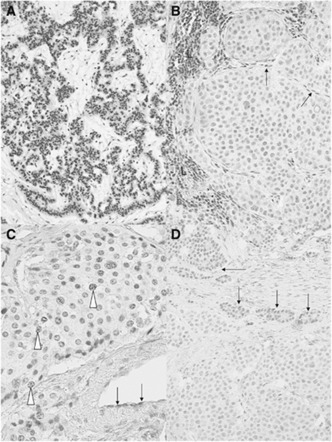
Figure 1.
Immunostaining for MGMT protein. Representative examples of strong positive nuclear staining of tumour cells in a pancreatic NET (A) and of unambiguous negative staining of an ileal NET (B) are shown. Note in B the nuclear staining of lymphoid cells (L) and endothelial cells (arrows), which serve as internal positive controls (B). (C) An example of non-interpretable staining in an ileal NET: despite a very faint positivity observed in some tumour cell nuclei (open arrowheads), the absence of any positive internal control, especially in endothelial cells (arrows), prevents the definitive interpretation of the case. In D, an example of highly heterogeneous pancreatic NET is shown, with two distinct tumour cell populations, one with a faint nuclear labelling (arrows) and the other with no detectable labelling. Immunoperoxidase with nuclear counterstaining using Mayer's haematoxylin. Original magnifications: A, × 120; B, × 350; C, × 380; D, × 240; scale bar=50 μm.

