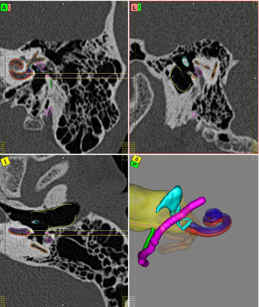
Figure 1.

Preoperative path planning. The results of the automatic segmentation and path planning are displayed to the surgeon for verification. Segmented structures include the scala tympani (red), scala vestibuli (blue), facial nerve (magenta), chorda tympani (green), ossicles (blue), and external auditory canal (yellow). The drill path is shown as a golden cylinder. Red line in the three-dimensional rendering shows the trajectory line.
