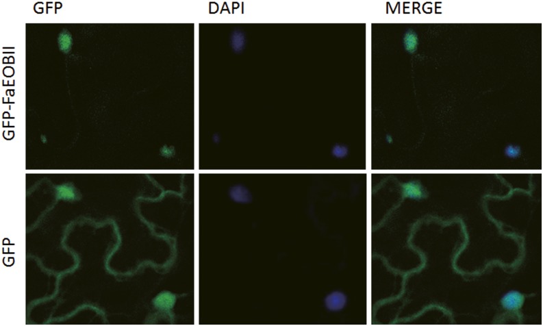
Figure 1.
Nuclear location of the FaEOBII protein in plant cells. Fluorescence signal was detected using a confocal microscope from GFP-FaEOBII (top row) and GFP (bottom row) expression under the control of the 35S promoter in N. benthamiana leaf epidermis cells. The MERGE column shows merged views of the GFP and DAPI images.

