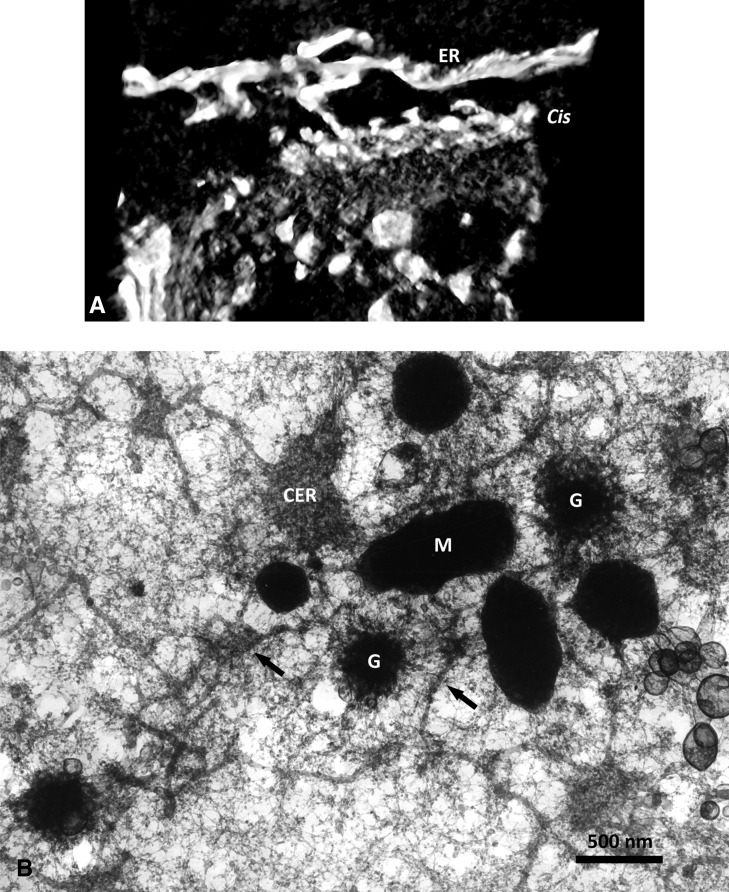
Figure 3.
A, Maximum-intensity projection in negative contrast of a stack of thin sections from a tomogram of a pea (Pisum sativum) root tip Golgi body and associated ER impregnated by the osmium zinc iodide technique. The reconstruction is presented at an angle to show a clear tubular connection between the ER and cis-Golgi. B, Inside face view of a dry-cleaved carrot (Daucus carota) suspension culture cell. The cell had been fixed on a coated EM grid, dehydrated, and critical point dried prior to dry cleaving on double-sided tape. The view onto the plasma membrane shows dark mitochondria (M), complete Golgi stacks in face view (G), cisternal ER (CER), and tubular ER (arrows). Note the huge difference between the diameter of a Golgi body and ER tubules.

