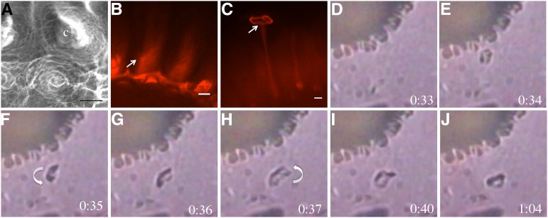
Figure 8.
Cellulose and pectin are organized in a spiral around the columella, which uncoils as mucilage expands when hydrated. A, VPSEM of hydrated mucilage. Cell wall material is oriented in spiral structures around the columella (c). B, S4B-stained ixr1-1 mucilage showing the spiraled orientation of cellulose fibrils in rays. The arrow indicates the orientation of the cellulose spiral. C, Maximum projection of a z-stack of S4B-stained fly1-1 seeds. The arrow indicates the point of ray attachment to the disc. Bars = 10 μm. D to J, fly1-1 Seeds hydrated in water, showing fly1 disc rotation during mucilage expansion. Times indicate seconds post hydration. For a clearer image of disc rotation, see Supplemental Video S3. Arrows indicate the direction of rotation.

