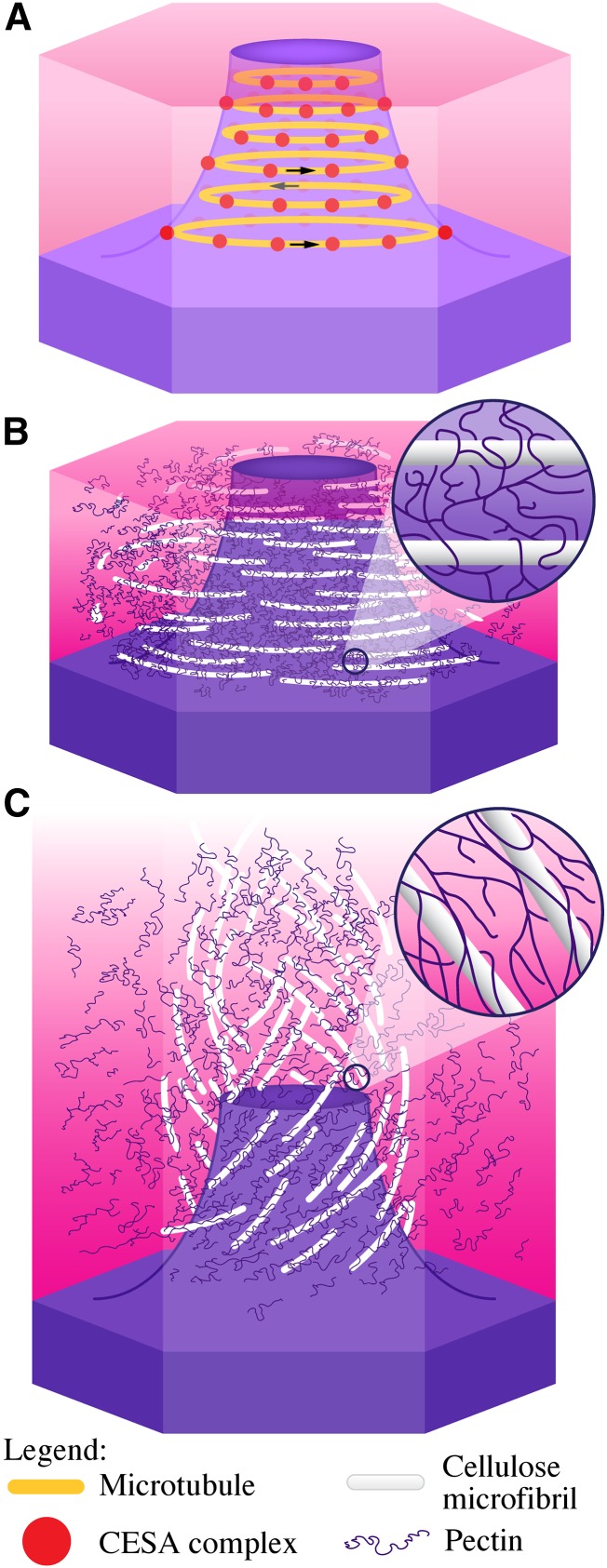
Figure 9.
Model of the orientation of cellulose deposition during mucilage biosynthesis and of mucilage expansion. A, Model of a seed coat epidermal cell at 7 DPA showing CESA complexes (red dots) trafficking around the cytoplasmic column circumferentially, guided by microtubules (orange lines). The direction of CESA complex movement is indicated by the arrows. B, Seed coat epidermal cell at 11 DPA showing cellulose and pectins in mucilage. The inset shows pectins wrapped around cellulose. C, Hydrated mucilage showing the orientation of cellulose microfibrils post hydration. The addition of water forces pectins to expand, pushing and pulling cellulose microfibrils in the process. The inset shows expanded pectins still wrapped around cellulose microfibrils.

