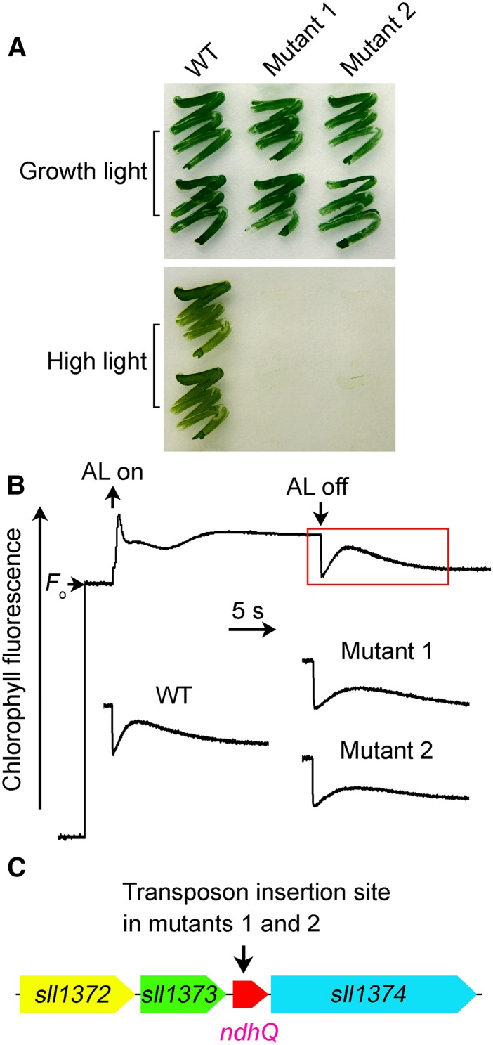
Figure 1.
Use of high light condition to screen for NDH-CET-defective mutant in transposon-tagged mutant populations of Synechocystis 6803. A, Growth of the wild type (WT) and mutants under normal light (40 µmol photons m−2 s−1) and high light (200 µmol photons m−2 s−1). B, Monitoring of NDH-CET activity using Chl fluorescence analysis. The top curve shows a typical trace of Chl fluorescence in the wild-type Synechocystis 6803. Cells were exposed to AL (620 nm; 45 µmol photons m−2 s−1) for 30 s. AL was turned off, and the subsequent change in the Chl fluorescence level was monitored as an indication of NDH-CET activity. C, The arrow schematically indicates the transposon insertion site in mutants 1 and 2 probed by PCR analysis using the primers listed in Supplemental Table S1.

