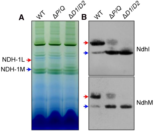
Figure 7.
Western analyses of NDH-1L and NDH-1M complexes from the wild-type (WT), ∆P/Q, and ∆D1/D2 strains. A, Thylakoid protein complexes isolated from the wild type and mutants were separated by BN-PAGE. Thylakoid membrane extract corresponding to 9 µg of Chl a was loaded onto each lane. The positions of the NDH-1L and NDH-1M complexes are indicated by red and blue arrows, respectively. B, Protein complexes were electroblotted to a polyvinylidene difluoride membrane, and the membrane was cross reacted with anti-NdhI and NdhM to probe assembly of the NDH-1L and NDH-1M complexes.

