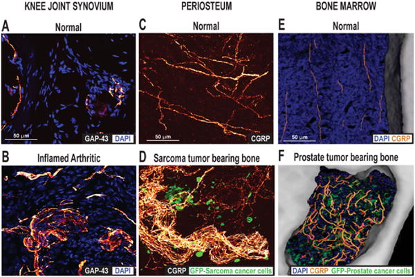
Figure 5.

Ectopic sprouting of primary afferent sensory nerve fibers occurs in a variety of skeletal pain conditions. (A) Confocal images of sections of (A) the normal and (B) the inflamed knee joint that have been immunostained for DAPI, which stains nuclei, and growth-associated protein (GAP-43), which stains sprouting nerve fibers. Twenty-eight days after the initial injection of Complete Freund's Adjuvant (CFA) into the rat knee-joint, a significant number of GAP-43+ nerve fibers in the synovial knee joint had sprouted and had a disorganized appearance, as compared with vehicle-injected mice. Reprinted with permission from Jimenez-Andrade & Mantyh (2012). Confocal images of periosteal whole mounts of (C) normal and (D) sarcoma tumor-bearing bone immunostained for calcitonin gene-related peptide (CGRP+) and green fluorescent protein (GFP)-labeled sarcoma. As tumor cells invade the periosteum of the bone, ectopic sprouting of CGRP+ sensory fibers occurs and neuroma-like structures form. Reprinted with permission from Mantyh et al. (2010). Confocal images of bone marrow of (E) normal and (F) prostate tumor-bearing bone marrow, immunostained for DAPI, CGRP and GFP-expressing prostate cancer cells. Note that in the normal mice, CGRP+ nerve fibers present in the marrow space of normal mice appear as single nerve fibers with a highly linear morphology. As GFP+ prostate tumor cells proliferate and form tumor colonies, the CGRP+ sensory nerve fibers undergo marked sprouting which produces a highly branched, disorganized and dense meshwork of sensory nerve fibers that is never observed in normal marrow. Reprinted with permission from Jimenez-Andrade et al. (2010).
