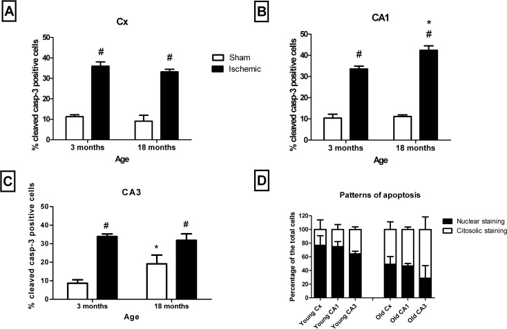
Fig. 6.
Age- and I/R-dependent apoptosis. Comparison of the percentage of apoptosis between young and old animals in the CX (a), CA1 (b) and CA3 (c) in sham-operated (open columns) and I/R-injured (black columns) animals. The pattern of staining in both young and old I/R-injured animals in the different structures is also shown (d). Age-dependent significant differences are represented by an asterisk, and I/R-dependent significant differences are represented by a number sign. No significant interactions between age and I/R were found (p < 0.05, two-way ANOVA, n = 5)

