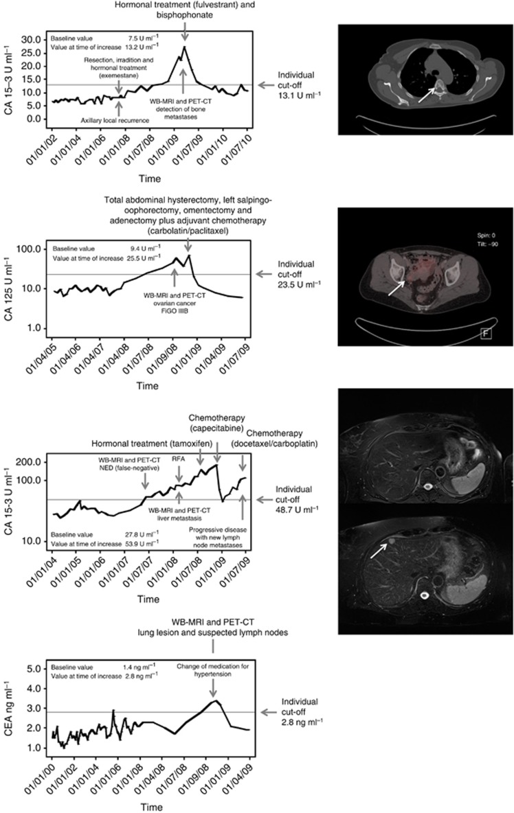
Figure 2.
Biochemical course of tumour markers and findings by whole-body imaging. (A) A 47-year-old patient with history of breast cancer (1998) and axillary local recurrence in June 2007. Metastases to the bone were detected in February 2009. At time of tumour marker increase, CT scan shows multiple bone metastases. One osteolytic metastasis is shown here at the fifth thoracic vertebral body. (B) A 53-year-old patient with history of breast cancer (2003) and primary diagnosis of ovarian cancer in September 2008. Fluorodeoxyglucose positron emission tomography/CT showed an increased uptake in the right ovary region susceptive of ovarian cancer. The patient was treated by resection and chemotherapy. The ovarian cancer was pathologically confirmed. (C) A 58-year-old patient with history of breast cancer (2001) and a tumour marker increase in July 2007, but no detectable malignancy whether in WB-MRI nor in FDG-PET/CT at the time of initial examination; liver metastasis were finally detected in follow-up imaging after 6 months. Whole-body-MRI at initial tumour marker increase did not show any morphologic suspected lesion in the whole body. After 6 months (control MRI), the axial T2-w fat saturated sequence shows a new focal metastatic lesion in segment 4a. Computed tomography-guided biopsy confirmed metastasis. (D) An 81-year-old patient with history of breast cancer (January 1995) and tumour marker increase in October 2009. In whole-body imaging, no correlate could be found. But, at time of tumour marker increase, the patient changed her medication for hypertension. After a while, CEA levels decreased to the individual baseline.

