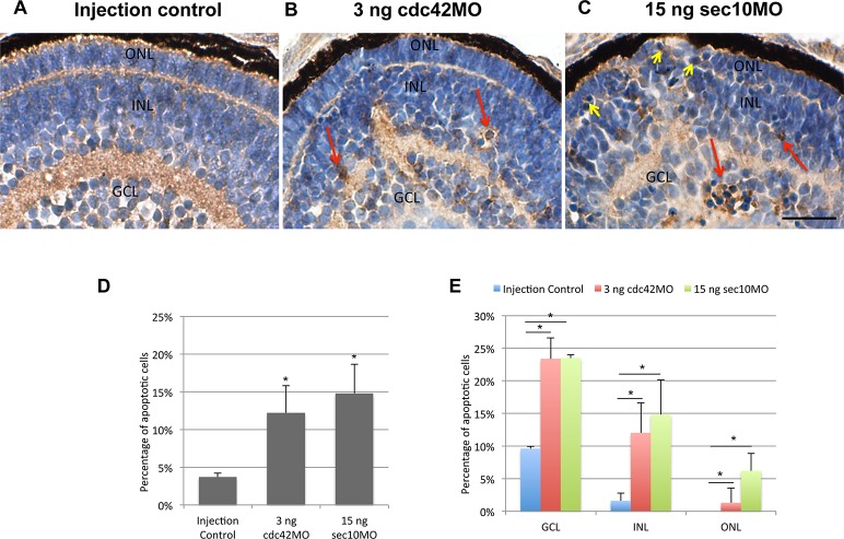
Figure 3.
Increased apoptosis is seen the retina of cdc42 and sec10 morphants at 72 hpf. (A–C) Casepase-3 staining reveals increased apoptotic cells within the retina of cdc42 and sec10 morphants. (D) The percentage of apoptotic retinal cells was significantly increased in cdc42 and sec10 morphants. (E) The frequency of apoptotic cells in the ONL, INL, and GCL of control, cdc42, and sec10 morphants is shown. *P < 0.001. Scale bars: 10 μm.

