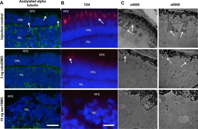
Figure 4.
The cdc42 and sec10 morphant embryos display abnormal OS development. (A, B) Immunofluorescence analysis of control, cdc42, and sec10 morphant zebrafish at 120 hpf. (A) Acetylated alpha tubulin (green), a marker for connecting cilia, localized to the OS region in control (arrow), but not cdc42 and sec10 morphants. (B) A marker for long double cones, 1D4 (red), localized to the OS region in control and cdc42 mutants. Staining was rarely observed in shorter OSs of cdc42 morphants (arrow), and no staining was observed in sec10 morphants. (C) Transmission electron micrographs of transverse sections along the dorsal-ventral axis of control, cdc42, and sec10 morphant zebrafish at 120 hpf. Low- and high-magnification images revealed longer OSs (arrow) in control embryos. Photoreceptor OSs were present in cdc42 morphants, but were significantly shorter and smaller in number than in controls. The OS was not observed in sec10 morphants. Scale bars: 10 μm.

