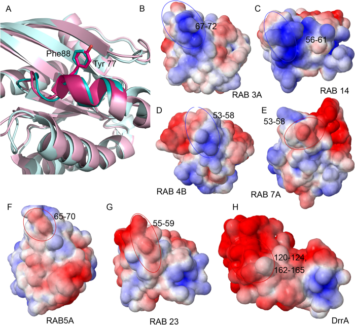
Figure 8. Substrate specificity of DrrA.
(A) Cartoon representation of Rab1b (PDB ID: 4HLQ, colored pink) and Rab 27a (PDB ID: 3BC1, colored blue). Stick representation depicts Tyr77 of Rab1b, which is AMPylated by DrrA and the structurally equivalent phenylalanine residue in Rab27a. (B-H) Electrostatic potential (±5 kT/e) rendered onto the surface of different Rab proteins and DrrA, positively charged surface is colored in blue and negatively charged surface in red. The potentials revealed positively charged surface in AMPylation compatible Rab proteins (highlighted in blue circles; B-D) and negative in AMPylation non-compatible Rab proteins (highlighted in red circles; E-G). (H) Negatively charged patch mapped on the molecular surface of DrrA.

