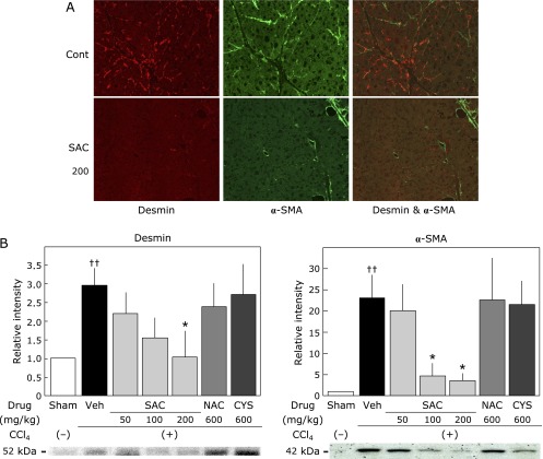
Fig. 7.
Effect of cysteine compounds on hepatic stellate cell activation in the liver. (A) Double immunofluorescence staining (anti-desmin labeled with FITC and anti-α-SMA labeled with PE) indicated a fine network pattern of stellate cells in the livers treated with CCl4 alone for 12 weeks (Control). Double-positive cells, presumably activated stellate cells, were observed in the control group. Conversely, desmin-positive (red) and α-SMA-positive (green) cells were hardly observed in SAC-treated groups (SAC 200). (B) Effect of cysteine compounds on the expression of desmin and α-SMA in the liver. Tissue lysates were evaluated by sodium dodecyl sulfate-polyacrylamide gel electrophoresis as described in the Materials and Methods. Animals were treated as described in the legend to Fig. 2. The relative increases of the intensity compared with the sham group are expressed as percentages. Values are the means ± SEM (n = 5–8). *p<0.05 vs the CCl4 group. ††p<0.01 vs the sham group. See online version figure.

