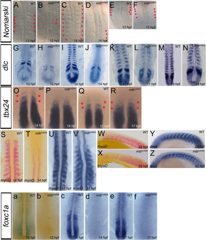
Figure 5.
Somite segmentation and specification recover without foxc1a expression. A–F are dorsal views of WT embryos and mibnn2002 mutants. Small round-shaped structures can be observed in (B) mibnn2002 homozygotes at 12 hpf. The newly-generated somite-like structures increased when mibnn2002 homozygotes grew to (D) 14 hpf and (F) 17 hpf. G–N are flat mounts of WT embryos and mibnn2002 mutants stained with dlc. (H) The expression of dlc in the region where somites normally formed was not observed in mibnn2002 homozygotes at 12 hpf. (J, L and N) dlc progressively appeared after 14 hpf in the newly-generated somites. O–R are dorsal views of WT embryos and mibnn2002 mutants stained with tbx24. The segmental tbx24 (marked with red asterisks) appeared progressively in mibnn2002 homozygotes. S–V are flat mounts of WT embryos and mibnn2002 mutants stained with myoD. W and X are lateral views of WT embryos and mibnn2002 mutants stained with myoD. The expression of myoD in formed somites started to emerge after 14 hpf as what was observed in dlc. As embryos developed, (V) the expression of myoD progressively appeared in the region where somites normally formed, except the anterior segments. (X) The cells that expressed myoD still split into dorsal and ventral myotomes even where somite boundaries were not formed. Y and Z are lateral views of WT embryos and mibnn2002 mutants stained with dystrophin (dmd) at 22 hpf. The expression of dystrophin can be observed in formed somites. Note that the somite gap delineated by dystrophin in formed somite region is similar between (Y) WT embryos and (Z) mibnn2002 mutants. a–f are dorsal views of WT embryos and mibnn2002 mutants stained with foxc1a. The expression of foxc1a in mibnn2002 homozygotes are not detected in all the stages examined (b 12 hpf; d 14 hpf and f 17 hpf).

