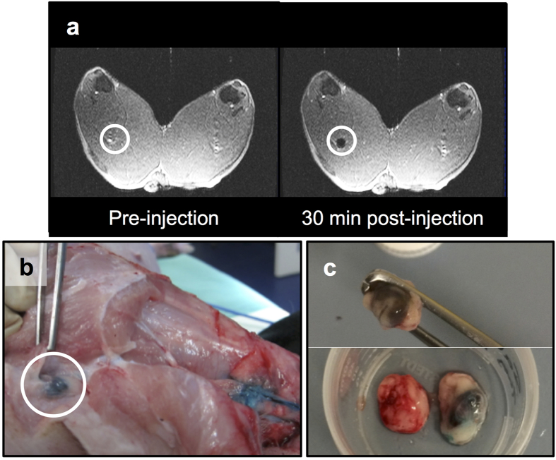
Figure 5. ‘Sentinel’ node identification in vivo.
(a) Preinjection and postinjection coronal MRI scans of a swine’s hind legs showing the negative contrast in one first-tier node (circled) resulting from the uptake of the magnetic tracer. Postinjection scans were used preoperatively to identify the location and number of first-tier nodes in both limbs for each animal. (b) After the anatomical location of the nodes had been determined with MRI, Patent Blue V dye was used to guide the surgery, and the identification of first-tier nodes (circled). (c) Some nodes were only visualised due to the dark brown staining from magnetic tracer accumulation (top). Compared to a control lymph node (bottom left), either blue dye (bottom right) or magnetic tracer uptake was sufficient to distinguish the nodes from surrounding tissue.

