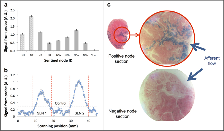
Figure 6. Swine lymph node measurements.
(a) MTJ magnetometer probe measurement of first-tier nodes from three animals. N1, N2, and control (Cont.) were measured ex vivo, and N3 – N6b were measured in vivo. Error bars indicate the uncertainty in measurement due to background signal fluctuations (electronic noise). (b) Distinguishing nodes in close proximity in ex vivo array of lymph nodes. The boundary of each node is indicated with the vertical dotted lines and the threshold level is indicated with the horizontal dotted line. (c) Prussian blue staining of two separate node sections. The presence of magnetic tracer in afferent trabecula and subcapsular sinuses is evident in positive first- and second-tier nodes (e.g. top), but absent in negative nodes (e.g. bottom).

