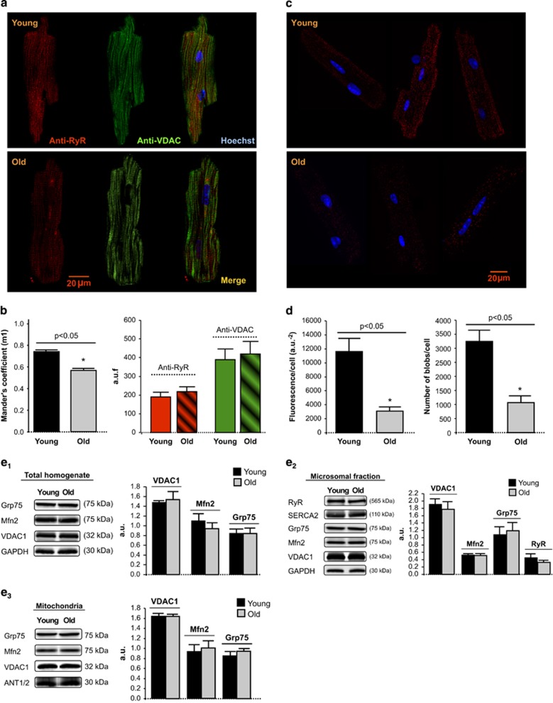
Figure 5.
(a) Confocal fluorescent images of young and old mouse cardiomyocyte simultaneously labeled with anti-RyR (red), anti-VDAC (green) and Hoechst (blue) for visualization of SR, mitochondria and nuclei, respectively. (b) Effect of aging on RyR–VDAC spatial interaction, as quantified by Mander's coefficient (m1) analysis, expressed as the percentage of RyR – with respect to total RyR fluorescence – that overlaps with VDAC (left panel); in the right panel, total RyR and VDAC fluorescence. Mean±S.E.M. from 4 to 6 cardiomyocytes per group (four hearts). (c) Confocal fluorescent images of the RyR–VDAC interaction in different individual cardiomyocytes isolated from young and old mouse hearts, detected by proximity ligation assay (PLA). Positive cross-reactivity – reflecting an intermolecular distance of <40nm – is shown in red, nuclei are depicted in blue (Hoechst). (d) Aging was associated with a significant reduction in cell fluorescence resulting from RyR–VDAC cross-reactivity (left panel) and in the number of amplification spots (right panel), as quantified by PLA assay. Mean±S.E.M. of 1071–3250 blobs per group (15 cardiomyocytes, two hearts). (e) Western blot representative bands and quantification of the expression of proteins involved in SR and mitochondria Ca2+ transport and interorganelle communication in whole-heart homogenate, microsomal and mitochondrial fractions from young and old mice. Each protein of interest was normalized by the corresponding protein of reference as follows: in total homogenates, VDAC1/Grp75, Mfn2/GAPDH and Grp75/GAPDH; in microsomal fraction, Mfn2/VDAC1, VDAC1/Grp75, Grp75/GADPH and RyR/Grp75; in mitochondria, VDAC1/Grp75, Grp75/ANT1/2 and Mfn2/ANT1/2. Mean±S.E.M. of n=8 replicates from four hearts/group)

