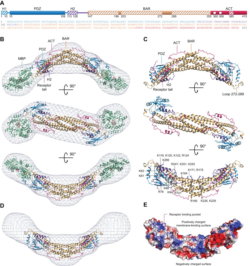
FIGURE 2:
Atomic model of PICK1 derived from SAXS. (A) Domain diagram and sequence of PICK1 (color coded). (B) Three perpendicular views of the average SAXS envelope fit with an atomic model of the MBP-PICK1 dimer (modeling details in text). PICK1 domains are color coded as in A, and MBP is shown in green. (C) PICK1 portion of the model extracted from the images shown in B. The third orientation shows the side chains of positively charged amino acids predicted to participate in membrane binding. (D) Same as B, but subtracting the MBP portion of the model to show the contribution of PICK1 to the overall envelope. (E) Electrostatic surface representation of the PICK1 model (red, negatively charged; blue, positively charged). See also Supplemental Movies S1–S3.

