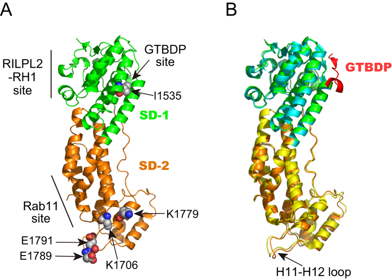
Figure 1. Structural comparison of Myo5a-GTD and Myo5a-GTD/Mlph-GTBDP.
(A) Ribbon representation of the human Myo5a-GTD structure (PDB ID: 4LX1) showing binding sites for Mlph-GTBDP, RILPL2-RH1, and Rab11. The residues mutated in our study are shown as spheres. (B) Overlap of the crystal structures of the human Myo5a-GTD (PDB ID: 4LX1) and Myo5a-GTD/Mlph-GTBDP complex (PDB ID: 4LX2). The overlap reveals a relatively large conformation change in the H11-H12 loop upon Mlph-GTBDP binding. Subdomain-1 (SD-1) and subdomain-2 (SD-2) are shown in green and orange for the apo-GTD and cyan and yellow for the GTD/Mlph-GTBDP complex.

