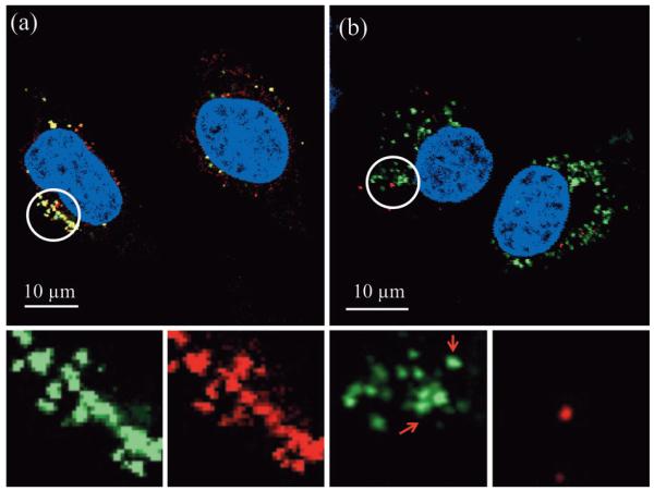
Figure 2.
Merged representative confocal fluorescent images of HeLa cells 24 h after incubation (≈3×10³ cells with 200 μg mL−1 of MSNs) with a) 5 mol% primary amine modified MSNs, type (2)b, and b) 1 mol% primary amine modified MSNs, type (2)a. Spectrally resolved components of the circled regions are magnified and shown below each image. The images shown are entirely representative of the behaviour observed across the population of cells for each given particle type. The yellow in (a) indicates co-localisation of MSNs and endo/lysosomes. This localisation is confirmed on unmixing the particle and lysomal emissions (note the near perfect overlap of green particles and red lysosomes). For 1 mol% particles (b), subcellular distribution is markedly different and there is very little alignment of particle and lysosome emission. The ability of these particular particles to access the cytosol is additionally confirmed by Z-stack analysis, additional location specific staining, and control experiments with different exterior chemistries and protein appendages (Figure S1 in the Supporting Information).

