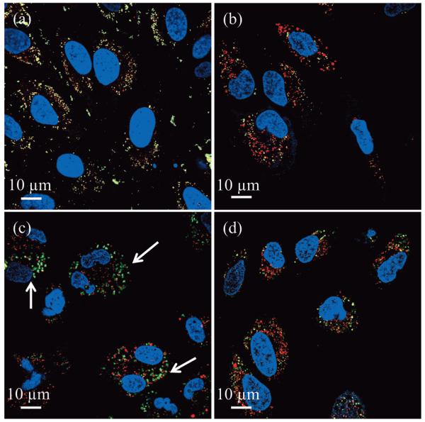
Figure 5.
Merged confocal fluorescent images of HeLa cells after 24 h incubation (≈3×10³ cells incubated with 200 μg mL−1 of MSNs) confirming that only 1 mol% primary amine and imidazole co-modified MSNs, type (4), are capable of escape into the cytosol. For the spectrally resolved components see Figure S3 in the Supporting Information. Images from (a) to (d) are the corresponding characteristic results associated with type (1), type (3), type (4) and type (5) particles, respectively. Yellow colour represents co-localised MSNs (green) and endo/lysosomes (red). Arrows indicate MSNs that have escaped endo/lysosomal entrapment. The images shown are entirely representative of the behaviour observed across the population of cells for each given particle type.

