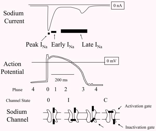
Fig. 1.
Peak, Early, and Late INa. Diagrams of cardiac sodium current (INa) action potential (AP), and sodium channel cartoon, lined up with time on the horizontal. Peak INa occurs in less than a millisecond and underlies the rapid upstroke or phase 0 of the AP. At this time, the activation gate and the inactivation gate on the channel are both open. Then over several milliseconds the current begins to decay contributing to a notch in the AP called phase 1. At this time, some of the channels have inactivated (shown as “I” with the inactivation gate closed). There is no commonly accepted name for this phase of INa but here it is labeled “Early INa”. After several milliseconds INa normally decays to <1% of peak INa, but a residual current flows as late INa and this depolarizing current along with calcium currents support phase 2 or the plateau of the AP. The mechanisms for late INa at the sodium channel level are multiple but can generally be thought of as incomplete inactivation. Eventually, activating potassium currents repolarize the membrane (phase 3 of the AP) and when the voltage decreases below the sodium channel threshold, the activation gate closes.

