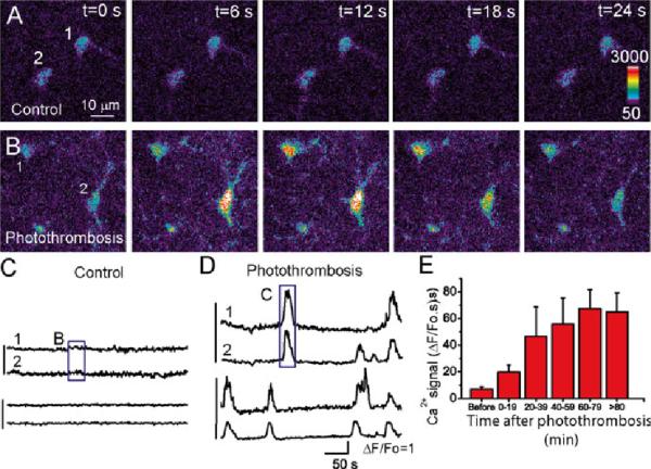
Fig. 10.4.
Enhanced Ca2+ signaling in astrocytes in the mouse cortex in vivo after photothrombosis. (a, b) Represent images of astrocytes loaded with ftuo-4 before (a) and after (b) photothrombosis. (c, d) Time courses of somatic Ca2+ oscillation of individual astrocytes expressed as ΔF/Fo before (c) and after photothrombosis (d). The box regions correspond to the images in (a) and (b) as indicated. (e) Time course of somatic Ca2+ signal in astrocytes developed after photothrombosis. Adapted from Ding et al. (2009)

