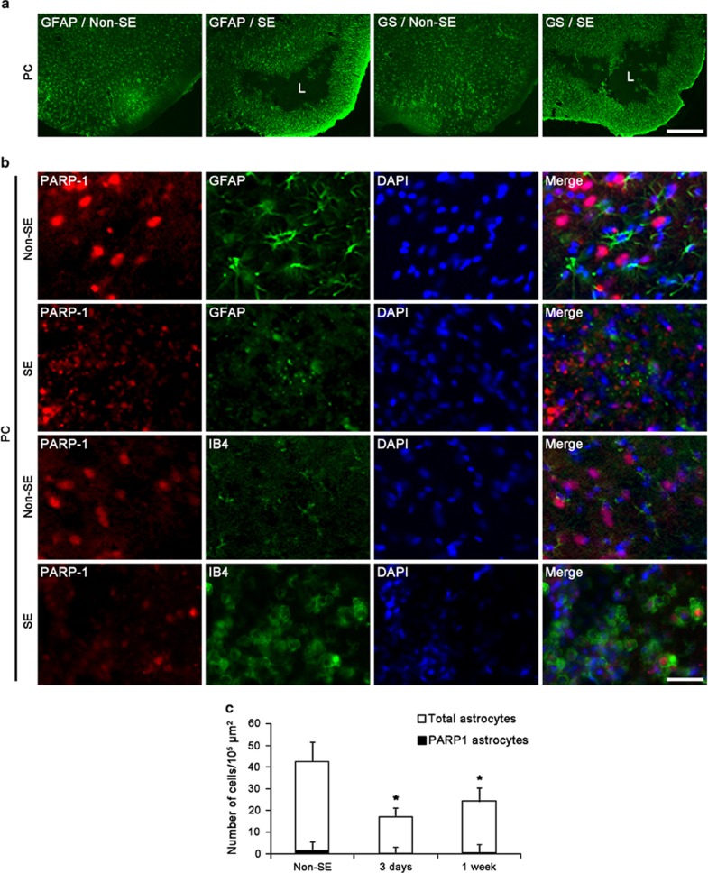
Figure 4.
Astroglial death and microglial PARP1 induction in the PC following SE. (a) Astroglial death induced by SE. Both GFAP and GS expression are reduced in the PC. Astroglial depleted lesion (L) is surrounded by reactive astrocytes. Bar=200 μm. (b) Double immunofluorescent images for PARP1 and GFAP/IB4 in the PC. In non-SE animals, PARP1 expression is detected only in PC neurons. Following SE, PARP1 degradation and GFAP-positive astroglial loss are observed in the PC. In addition, IB4-positive microglia show PARP1 immunoreactivity. Bar=12.5 μm. (c) The changes in the number of total astrocytes and PARP1-positive astrocytes in the hippocampus following SE. The number of total astrocytes is reduced at 3 days to 1 week after SE, whereas the fraction of PARP1-positive astrocytes in total astrocytes is unaltered (means±S.D., n=5, respectively). *P<0.05 versus non-SE animals (one-way ANOVA test)

