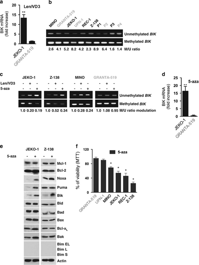
Figure 6.
The Len/VD3 treatment increased BIK mRNA expression via the demethylation of CpG islands. (a) The Len/VD3 treatment increased BIK mRNA expression. JEKO-1 cells were treated for 4 days with 1 μM Len and 100 nM VD3. BIK mRNA expression was quantified by qRT-PCR assay. The data represent the mean±S.E. of three independent experiments. (b) BIK CpG islands were constitutively methylated in MCL cell lines and patient samples (P1–P4). Methylation-specific PCR was performed on genomic DNA as described within the Material and Methods section and in Supplementary Figure S3. (c) The Len/VD3 treatment induced the demethylation of BIK CpG islands. MCL cells were treated for 4 days with 1 μM Len and 100 nM VD3 or for 3 days with 1 μM 5-aza. Methylation-specific PCR was performed on genomic DNA. (d) 5-aza increased expression of BIK mRNA. MCL cells were treated for 3 days with 1 μM 5-aza. qRT-PCR assay was performed as described within the legend of Figure 6a. The data represent the mean±S.E. of three independent experiments. (e) 5-aza induced expression of Bik. MCL cells (2 × 10⁵/ml) were incubated for 3 days with or without 1 μM 5-aza. Cells were then lysed and expression of the indicated proteins was assessed by western blotting. (f) 5-aza induced cell death in MCL cell lines sensitive to Len/VD3 treatment. Cells (2 × 10⁵/ml) were treated for 3 days with 1 μM 5-aza and cell viability was assessed using MTT assay. The data represent the mean±S.E. of three independent experiments. *P<0.05, **P<0.01

