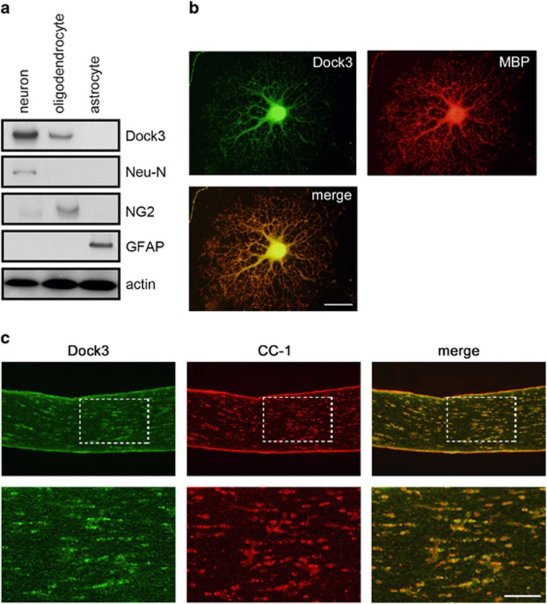
Figure 1.
Expression of Dock3 in oligodendrocytes. (a) Expression of Dock3 and markers for neurons (Neu-N), oligodendrocytes (NG2) and astrocytes (GFAP) in cultured cells prepared from mouse brain. Note the expression of Dock3 in oligodendrocytes as well as in neurons. (b and c) Double-labeling immunohistochemistry for Dock3 and myelin markers in cultured oligodendrocyte (b) and optic nerve (c). Scale bar: 20 μm in b and 100 and 50 μm in the upper and lower panels in c

