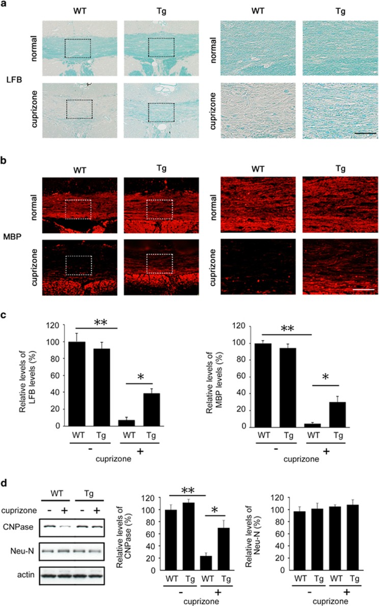
Figure 2.
Effects of Dock3 on myelin protection in the corpus callosum of a cuprizone-induced demyelinating model. (a and b) Brain coronal sections illustrating a high intensity of LFB (a) and MBP (b) in Dock3 Tg mice compared with WT mice after cuprizone treatment for 6 weeks. Right panels are higher magnifications of the boxes shown in the left panels. Scale bars: 150 μm in the left panels and 50 μm in the right panels. (c) Quantitative analysis of LFB- and MBP-intensities in a and b. (d) Western blot analysis displaying increased CNPase expression in the brain of Dock3 Tg mice after cuprizone treatment. The data are presented as mean±S.E.M. of six independent samples. The results are expressed as percentages of the normal WT mice. **P<0.01; *P<0.05

