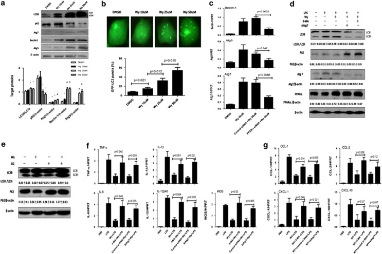
Figure 6.
PPARα activation suppresses proinflammatory response by promoting autophagy in vitro. (a) BMDMs were stimulated with or without Wy-14 643 (10, 25 or 50 μM). Total cell lysates were analyzed for LC3B, p62, Atg7, Beclin-1 and Atg5, as well as total β-actin protein levels by western blotting. Densitometry analysis of the proteins was performed for each sample. Data are shown as mean±S.E.M (n=3). *P<0.05 or #P<0.01, compared with dimethylsulfoxide (DMSO)-treated BMDMs. (b) Transfected GFP-LC3 plasmid for 12 h, BMDMs were preincubated with Wy-14 643 (10, 25 or 50 μm) for 24 h to observe the formation of autophagosomes. The percentage of cells with GFP-LC3 puncta in the groups receiving different concentrations. GFP-positive cells were defined as cells that display bright, punctate staining. Approximately 50 cells were counted, and the experiment was repeated at least three times. (c) After transfection of PPARα siRNA or control siRNA (3 mg/ml) for 36 h, BMDMs were incubated with or without Wy-14 643 (50 μM) for 6 h. Gene induction of Atg7, Atg5 and Beclin-1 was measured by qRT-PCR. The average target gene/HPRT ratios for each experimental group were plotted. (d) After transfection of Atg7 siRNA (3 mg/ml) for 36 h or preincubation with 3-MA (10 mM/l) for 2 h, BMDMs were incubated with or without Wy-14 643 (50 μM) for 2 h and then incubated in LPS (20 ng/ml) for 6 h. Western blot analysis shows the LC3BII/I ratio, Atg7, PPARα and p62 degradation. β-Actin is shown as a loading control; densitometry analysis of the proteins was performed for each sample; data are shown as mean±S.E.M (n=3). (e) Wy-14 643 activates autophagic flux in the primary hepatocytes of mice. Hepatocytes were treated with 50 μM Wy-14 643 in the absence or presence of CQ (10 μM) for 24 h. Western blot analysis shows the LC3BII/I ratio and p62 degradation. β-Actin is shown as a loading control; densitometry analysis of the proteins was performed for each sample; data are shown as mean±S.E.M (n=3). (f and g) After transfection of Atg7 siRNA (3 mg/ml) for 36 h or preincubated 3-MA (10 mM/l) for 2 h, BMDMs were incubated with or without Wy-14 643 (50 μM) for 2 h and then incubated with LPS (20 ng/ml) for 6 h. Cytokine and chemokine gene induction was measured by qRT-PCR. The average target gene/HPRT ratios for each experimental group were plotted

