
Figure 2. Narrow time window and stringent dose response to RA for development of functional mucosal-like DCs.
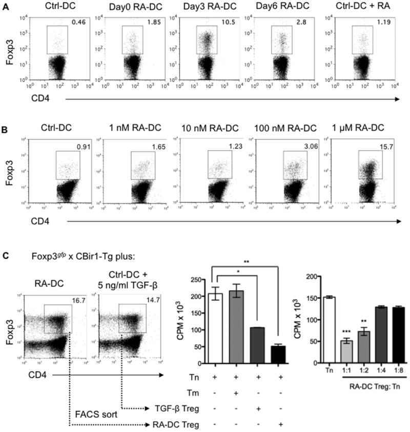
(A) BMDCs generated as described in Figure 1 were pulsed with CBir1 flagellin peptide and then cocultured with FACS-sorted CD4+Foxp3gfp- naïve T cells from Foxp3gfp.CBir1-Tg mice. T cell Foxp3 expression was examined by flow cytometry after 5 days. (B) Doses from 1 nM to 1 μM of RA were added to BMDC cultures from day 3. On day 8, BMDCs pulsed with CBir1 flagellin peptide were cocultured with FACS-sorted CD4+Foxp3gfp- CBir1-Tg naïve T cells. T cell Foxp3 expression was examined by flow cytometry after 5 days. Plots are gated on CD4+ cells, and the percentages of Foxp3+ cells are shown (A, B). (C) Naïve Foxp3gfp.CBir1-Tg CD4+ T cells were cultured with day3 RA-DCs, or with control BMDCs in the presence of 5 ng/ml of TGF-β. CD4+Foxp3gfp+ T cells were sorted by FACS 5 days later. Naïve CBir1-Tg CD4+ T cells were cultured alone (Tn), with memory T cells (Tm), or with FACS-sorted Foxp3gfp+ cells from both conditions at a ratio of 1:1 or as indicated in the presence of CBir1 flagellin peptide-pulsed splenic feeder cells. Tritiated thymidine was added for the last 18 h of a 72-h incubation period for T cell proliferation. Mean CPM of triplicates ± SEM are shown (*, P < 0.05; **, P < 0.01; ***, P < 0.001 as compared to Tn). Data are representative of three experiments.