
Figure 3. TGF-β and RA differentially regulate RA-DC induction of CD4+ T cell Foxp3 and CCR9 expression.
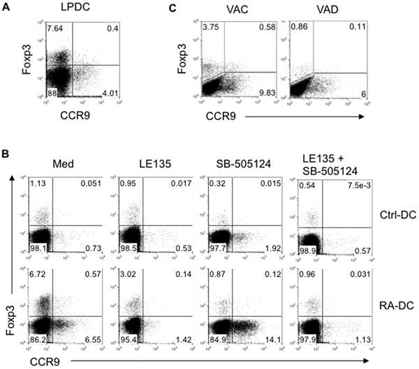
(A) LP-DCs were isolated from C57BL/6 mice and cultured with naïve CBir1-Tg CD4+ T cells in the presence of CBir1 flagellin peptide. Expression of Foxp3 and CCR9 by CD4+ T cells was determined by flow cytometry after 5 days. (B) CBir1 flagellin peptide-pulsed control BMDCs and day3 RA-DCs were cultured with naïve CBir1-Tg CD4+ T cells in the presence or absence of TGF-β RI inhibitor SB-505124, and/or RAR antagonist LE135. Expression of Foxp3 and CCR9 by CD4+ T cells was determined by flow cytometry 5 days later. (C) MLN DCs isolated from control (VAC) or vitamin A-deficient (VAD) mice were pulsed with CBir1 flagellin peptide and cocultured with naïve CBir1-Tg CD4+ T cells. Expression of Foxp3 and CCR9 by CD4+ T cells was determined 5 days later by flow cytometry. Data are representative of two (C) or three (A, B) experiments.